Introduction to Animal Cell Coloring Pages
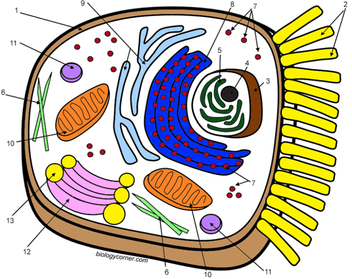
Coloring page for animal cell – Unlocking the wonders of biology can be an exciting adventure, especially for young minds. Animal cell coloring pages offer a unique and engaging way to learn about the fundamental building blocks of life. These vibrant activities transform the often-abstract concept of the cell into a fun and memorable experience, fostering a deeper understanding of biological structures and processes.Coloring animal cells is beneficial for children of all ages, sparking curiosity and promoting active learning.
The simplicity of the activity makes it accessible to preschoolers, while the complexity of cellular structures provides a stimulating challenge for older children.
Educational Value and Age Appropriateness
Animal cell coloring pages offer a multi-faceted approach to learning. Younger children, from preschool to early elementary school, can focus on basic shapes and colors, learning to identify the major components of an animal cell like the nucleus and cell membrane. Older children, from late elementary school through middle school, can delve into more detailed representations, incorporating organelles such as mitochondria, ribosomes, and the Golgi apparatus.
This graduated approach ensures that the activity remains both engaging and appropriately challenging for different developmental stages.
Learning Objectives Achieved Through Coloring
Coloring activities go beyond simple entertainment; they contribute significantly to cognitive development. Through coloring animal cells, children enhance their fine motor skills, improving hand-eye coordination and dexterity. The process of identifying and coloring different organelles reinforces memory and recall of scientific terminology. Furthermore, the act of creating a visual representation of an abstract concept strengthens comprehension and allows for a deeper, more intuitive understanding of animal cell structure and function.
For example, coloring the nucleus a distinct color helps children associate that specific shape and color with the cell’s control center. Similarly, distinguishing the mitochondria with a different hue helps them understand its role in energy production. The visual association created through coloring significantly improves retention and understanding.
Design Aspects of Animal Cell Coloring Pages
Creating engaging and educational animal cell coloring pages requires careful consideration of design elements to effectively convey complex biological information in a visually appealing and accessible manner. The goal is to make learning about cell structures and processes fun and memorable for children and students of all ages. Well-designed coloring pages can foster a deeper understanding of the intricacies of animal cells, turning a potentially dry subject into an enjoyable activity.
Effective design incorporates clear visuals, accurate representations of organelles, and engaging layouts that stimulate creativity and learning. The use of color and labeling can greatly enhance understanding and retention of information. Moreover, different coloring page designs can be employed to target various learning objectives, such as understanding cell structure, cell division, or comparing animal and plant cells.
A Basic Animal Cell Coloring Page
This coloring page depicts a simplified animal cell, highlighting major organelles. The cell is presented as a roughly circular shape with a large, centrally located nucleus. The nucleus contains a smaller, darker circle representing the nucleolus. Scattered throughout the cytoplasm are numerous mitochondria, depicted as bean-shaped structures with internal cristae (folds) indicated by smaller, parallel lines within each mitochondrion.
Small dots represent ribosomes, dispersed throughout the cytoplasm. The cell membrane, a thin outer boundary, encloses the entire cell. A key is provided, identifying each organelle with its name and a corresponding color suggestion (e.g., nucleus – purple, mitochondria – red, ribosomes – blue, cytoplasm – yellow, cell membrane – light green). The page might include simple, clear labeling to further aid in identification.
Animal Cell Mitosis Coloring Page
This coloring page illustrates the stages of mitosis, the process of cell division in animal cells. The page could show a series of four or five panels, each representing a different phase: prophase, metaphase, anaphase, and telophase. Each panel depicts the cell at a specific stage, clearly showing the changes in chromosome arrangement and the behavior of the cell’s structures.
For example, in prophase, the chromosomes would be shown condensing and becoming visible, while in anaphase, the sister chromatids would be separating and moving to opposite poles of the cell. A clear key would identify each phase and the key cellular structures involved, such as chromosomes, spindle fibers, and centrioles. Simple, clear labels for each stage would be included to reinforce learning.
The final panel shows the two daughter cells resulting from successful mitosis.
Comparing Animal and Plant Cells
This coloring page focuses on the differences between animal and plant cells. It features side-by-side depictions of an animal cell and a plant cell, each with its key organelles clearly labeled. A table directly below the cell illustrations highlights the key differences.
| Feature | Animal Cell | Plant Cell | Difference Explained |
|---|---|---|---|
| Cell Wall | Absent | Present (rigid, made of cellulose) | Plant cells have a rigid outer layer for support and protection, while animal cells lack this structure. |
| Chloroplasts | Absent | Present (sites of photosynthesis) | Plant cells contain chloroplasts to carry out photosynthesis, a process absent in animal cells. |
| Vacuole | Small or absent | Large central vacuole | Plant cells usually have a large central vacuole for storage and turgor pressure; animal cells have smaller, less prominent vacuoles. |
| Shape | Variable (round, irregular) | Usually rectangular or polygonal | Plant cells have a more defined shape due to the cell wall; animal cells exhibit more flexibility in shape. |
Organelle Focus
Let’s delve into the fascinating world of animal cell organelles, bringing their intricate structures and vital functions to life through detailed illustrations. Understanding these tiny powerhouses is key to appreciating the complexity and wonder of the cell itself. This section provides detailed descriptions perfect for creating vibrant and informative coloring pages.
The Nucleus: Control Center of the Cell
The nucleus is the cell’s command center, a large, membrane-bound organelle containing the cell’s genetic material, DNA. Imagine it as a spherical or oval-shaped structure, easily recognizable by its prominent size within the cell. For your coloring page, depict it with a double membrane, the nuclear envelope, punctuated by nuclear pores – tiny openings that regulate the passage of molecules in and out.
Inside, illustrate the densely packed chromatin, which is DNA organized with proteins. You can represent this as a network of tangled threads or as more condensed regions called nucleoli, which are responsible for ribosome production. The nucleus dictates the cell’s activities by controlling gene expression, ultimately determining what proteins are made and when.
Mitochondria: The Powerhouses
Mitochondria are the energy factories of the cell, generating the energy currency known as ATP (adenosine triphosphate) through cellular respiration. Illustrate them as bean-shaped or sausage-shaped organelles with a double membrane. The outer membrane is smooth, while the inner membrane is highly folded, forming structures called cristae. These cristae significantly increase the surface area for ATP production.
For a visually engaging coloring page, depict the cristae as numerous folds within the inner membrane, highlighting their crucial role in energy generation. The space between the inner and outer membranes is called the intermembrane space, and the space inside the inner membrane is the mitochondrial matrix. The matrix is where many of the reactions of cellular respiration take place.
Endoplasmic Reticulum: The Cellular Highway System
The endoplasmic reticulum (ER) is a network of interconnected membranes extending throughout the cytoplasm. There are two types: rough ER and smooth ER. The rough ER, so named for its studded appearance, is dotted with ribosomes, the protein synthesis machinery. Illustrate the rough ER as a series of interconnected flattened sacs or cisternae, with small dots representing ribosomes attached to its surface.
The ribosomes synthesize proteins, which then enter the lumen (interior space) of the rough ER for folding and modification. The smooth ER, lacking ribosomes, plays a key role in lipid metabolism, detoxification, and calcium storage. Depict the smooth ER as a network of interconnected tubules, distinct from the flattened sacs of the rough ER. Its smooth appearance reflects the absence of ribosomes.
These two types of ER work together, with proteins synthesized on the rough ER often moving to the smooth ER for further processing before being transported to their final destinations.
Activity and Learning Extensions: Coloring Page For Animal Cell
Unlocking a deeper understanding of the animal cell goes beyond simply coloring. Engaging activities can transform the coloring page into a springboard for creative exploration and knowledge retention. These activities encourage active learning, fostering a more comprehensive grasp of cell structure and function.The following activities and lesson plan integrations offer diverse pathways for students to explore the intricacies of the animal cell, turning a simple coloring exercise into a memorable and impactful learning experience.
By combining visual learning with hands-on activities and storytelling, we can ignite curiosity and cultivate a lasting appreciation for the wonders of biology.
Creative Activities to Enhance Learning
These five activities build upon the visual learning provided by the coloring page, encouraging students to engage with the material in diverse and creative ways. Each activity promotes a different learning style, ensuring broad accessibility and engagement.
- 3D Animal Cell Model Construction: Students can use various materials like clay, foam balls, pipe cleaners, and construction paper to build a three-dimensional model of an animal cell, referencing their colored page for accuracy in organelle placement and size. This hands-on activity solidifies their understanding of spatial relationships between organelles.
- Animal Cell Comic Strip Creation: Students can create a comic strip depicting the daily life of an animal cell, personifying organelles and illustrating their functions in a fun and engaging way. This exercise promotes understanding of organelle interaction and collaboration.
- Animal Cell Acrostic Poem: Students can write an acrostic poem using the letters of “Animal Cell,” with each line describing a different organelle or its function. This activity encourages creative writing and reinforces vocabulary related to cell biology.
- Animal Cell Role-Playing: Students can role-play as different organelles within an animal cell, acting out their individual functions and interactions. This activity enhances understanding of collaborative processes within the cell.
- Design Your Own Animal Cell: Students can design their own hypothetical animal cell, adding or modifying organelles and explaining the rationale behind their design choices. This activity fosters critical thinking and encourages exploration of cellular adaptations.
Lesson Plan Integrations
Integrating the coloring pages into existing lesson plans provides a visual anchor for learning, making abstract concepts more concrete and memorable for students. These scenarios demonstrate versatile applications in different learning environments.
- Introduction to Cell Biology: The coloring page can serve as an introductory activity, allowing students to familiarize themselves with the basic structure of an animal cell before delving into more complex concepts. Following the coloring activity, students can engage in the 3D model construction activity mentioned previously.
- Organelle Function Exploration: After a lecture on specific organelles and their functions, students can use the coloring page to reinforce their learning by labeling and coloring the organelles. The Animal Cell Acrostic Poem activity would be a suitable follow-up.
- Cellular Processes and Interactions: Following a lesson on cellular processes like respiration or protein synthesis, students can use the coloring page to visualize the organelles involved. The Animal Cell Role-Playing activity would be an effective method to consolidate their understanding.
A Short Story: The Adventures of Cellula
Cellula, a spirited mitochondrion, lived in the bustling city of Cellville. Her job was to generate energy for the entire city, a vital task for keeping everything running smoothly. One day, a rogue virus, disguised as a friendly protein, tried to infiltrate Cellville. Cellula, along with her friends the ribosomes (who made the city’s defenses) and the lysosomes (the city’s sanitation workers), worked together to fight off the virus.
The nucleus, the city’s wise mayor, provided strategic guidance. Through teamwork and efficient processes, they repelled the virus and Cellville thrived. This adventure highlights the interconnectedness and importance of each organelle within the cell.
Creating a coloring page for an animal cell offers a unique opportunity to learn about biology in a fun way. The intricate details of the cell’s organelles can be engaging, much like the vibrant details found in coloring images of wild animals , which also offer a fantastic way to explore the natural world. Returning to the animal cell, remember to color the nucleus and cytoplasm with care, representing the bustling activity within!
Accessibility and Inclusivity
Creating accessible and inclusive animal cell coloring pages ensures that all students, regardless of their abilities or learning styles, can participate and benefit from this engaging learning activity. By thoughtfully designing and adapting the pages, we can foster a truly inclusive classroom environment where every student feels empowered to learn and express their creativity.Designing inclusive coloring pages requires careful consideration of various factors to ensure that they are usable and enjoyable for all learners.
This involves simplifying designs for younger children or those with specific learning needs, adapting the pages for visual impairments, and catering to diverse learning styles. The goal is to create a learning experience that is both effective and enjoyable for every student.
Simplified Illustrations for Diverse Learners
Simplified illustrations are crucial for accessibility. Younger children and students with certain learning differences may struggle with intricate details. Consider using bolder Artikels, larger organelles, and fewer overlapping elements. For example, instead of depicting a complex Golgi apparatus with numerous cisternae, a simplified representation with only a few clearly defined layers would be more appropriate. The mitochondria could be represented as simple bean shapes instead of intricate, folded structures.
This approach allows for easier coloring and comprehension, making the activity more manageable and less frustrating. Using bright, contrasting colors for different organelles will further enhance visibility and understanding.
Adaptations for Visually Impaired Students, Coloring page for animal cell
For students with visual impairments, several adaptations can significantly enhance the coloring page experience. Consider using raised-line drawings or tactile materials to create a three-dimensional representation of the cell. Thick, textured lines allow for easier tracing and coloring with different tools. Alternatively, providing large-print versions or digital versions with adjustable font sizes and color contrast can be beneficial.
Describing the organelles in detail, using tactile representations such as textured paper or raised stickers corresponding to each organelle, offers an alternative way for students to interact with and learn about the cell structure. Audio descriptions of the cell and its components can further enhance understanding and engagement for visually impaired students.
Catering to Diverse Learning Styles
Different learning styles necessitate varied approaches to the coloring page activity. For visual learners, the coloring page itself serves as an excellent tool. For kinesthetic learners, incorporating tactile elements like textured paper or modeling clay to represent organelles can significantly enhance engagement. Auditory learners can benefit from audio descriptions of the organelles and their functions, alongside the visual representation.
Providing a variety of coloring tools—crayons, markers, colored pencils—allows students to choose the tools that best suit their preferences and skills. Furthermore, incorporating interactive elements such as matching games or quizzes related to the organelles can further cater to diverse learning styles and make the learning process more interactive and fun.
Coloring Page Examples
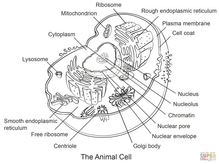
Let’s explore some examples of animal cell coloring pages, showcasing varying levels of complexity to cater to different age groups and learning styles. These examples illustrate how detail and color can enhance understanding of this fundamental unit of life. Each design offers a unique opportunity to engage with the intricate workings of the animal cell.
The following coloring pages demonstrate the progressive complexity of representing an animal cell, from a simplified overview suitable for younger learners to a more detailed representation for older students.
Simple Animal Cell Coloring Page
This coloring page provides a basic introduction to the major components of an animal cell. It’s ideal for younger children or as a starting point for those new to cell biology.
- Cell Membrane
- Cytoplasm
- Nucleus
A simple color palette could use light blue for the cytoplasm, dark blue for the nucleus, and a light brown for the cell membrane. This provides a clear visual distinction between the major components without overwhelming the learner.
Intermediate Animal Cell Coloring Page
This coloring page introduces more organelles and allows for a more detailed representation of the cell’s structure. It’s suitable for older elementary or middle school students.
- Cell Membrane
- Cytoplasm
- Nucleus (with nucleolus)
- Mitochondria
- Ribosomes
- Endoplasmic Reticulum (rough and smooth)
A more advanced palette could use different shades of green for the cytoplasm and organelles, representing the various metabolic processes occurring within the cell. For instance, darker greens for the mitochondria (energy production) and lighter greens for the endoplasmic reticulum (protein synthesis and lipid metabolism). The nucleus could be a deeper blue, while the nucleolus is a lighter shade. Ribosomes could be represented by tiny purple dots.
Advanced Animal Cell Coloring Page
This coloring page offers the most detailed representation, including a wider array of organelles and their interrelationships. This is appropriate for high school students or those with a deeper interest in cell biology.
- Cell Membrane
- Cytoplasm
- Nucleus (with nucleolus and chromatin)
- Mitochondria
- Ribosomes
- Endoplasmic Reticulum (rough and smooth)
- Golgi Apparatus
- Lysosomes
- Centrioles
- Vacuoles
For this level, a vibrant and diverse palette can highlight the specialized functions of each organelle. Mitochondria could be a deep red, reflecting their energy-producing role. The Golgi apparatus could be a bright orange, signifying its role in packaging and transporting molecules. Lysosomes, with their digestive function, could be a dark purple. This allows for a visually rich representation of the cell’s complexity.
Coloring Page Depicting the Cell Membrane and its Functions
This coloring page focuses specifically on the cell membrane, emphasizing its crucial role in selective permeability. The design should illustrate the phospholipid bilayer and embedded proteins, highlighting how different molecules interact with the membrane.
The page should depict various molecules attempting to cross the membrane: some successfully passing through (e.g., small, nonpolar molecules), others requiring assistance from transport proteins (e.g., larger polar molecules or ions), and others being blocked entirely (e.g., large molecules). The accompanying text could explain the concepts of diffusion, osmosis, and facilitated diffusion.
The cell membrane is selectively permeable, meaning it regulates what enters and exits the cell. This control is vital for maintaining the cell’s internal environment and its proper functioning. The illustration should clearly show how this selective permeability is achieved through the structure of the membrane and the actions of transport proteins.

