Answer Key Development

Animal cell coloring sheet answer key pdf – Creating a comprehensive and user-friendly answer key for an animal cell coloring sheet is crucial for effective assessment and learning. A well-designed answer key allows for quick and accurate grading, providing students with immediate feedback on their understanding of cell structures and their locations. This facilitates both self-assessment and teacher assessment, enhancing the overall learning experience.The development of the answer key should mirror the complexity of the coloring sheet itself.
Clear, concise labeling and a consistent format are essential for ease of use. The key should not only identify organelles but also clearly indicate their positions within the cell diagram.
Answer Key Formats
Several formats can be used to create an effective answer key. The choice depends on the specific needs and preferences of the educator and the complexity of the coloring sheet. A simple approach might involve numbering the organelles and providing a corresponding list with names and locations. Alternatively, letters or color codes could be used for identification.
- Numerical Format: This approach assigns a number to each organelle on the coloring sheet. The answer key then provides a numbered list matching the numbers to the organelle names and locations (e.g., 1. Nucleus – centrally located; 2. Mitochondria – scattered throughout the cytoplasm; etc.).
- Alphabetical Format: Similar to the numerical format, this uses letters instead of numbers. Each organelle is assigned a letter on the coloring sheet, and the answer key provides a corresponding alphabetical list (e.g., A. Nucleus – centrally located; B. Mitochondria – scattered throughout the cytoplasm; etc.).
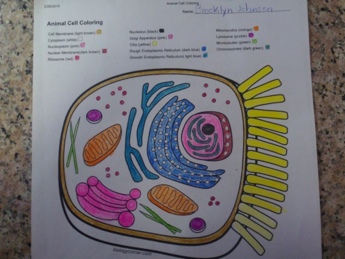
- Color-Coded Format: This method uses different colors to represent different organelles. The coloring sheet itself might use a legend to indicate which color represents which organelle. The answer key would then simply list the organelle name and its corresponding color.
Example Answer Key (Numerical Format)
This example uses a numerical format and assumes a simplified animal cell diagram.
Finding an animal cell coloring sheet answer key pdf can be really helpful for checking your work and understanding the different organelles. However, to truly grasp the concept, consider exploring the various ways you can color each part; for instance, check out this resource on animal cell coloring different colour for inspiration. Using different colors can improve memorization, ultimately making the answer key even more useful in reinforcing your learning.
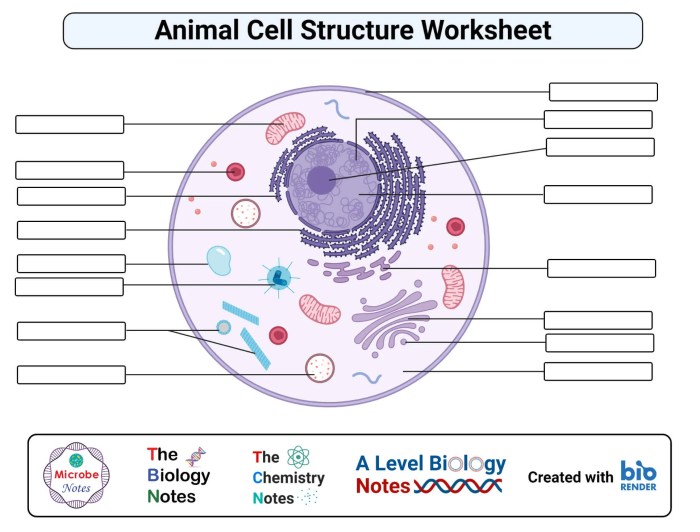
- Nucleus (1): Located centrally within the cell, this is the control center containing the cell’s genetic material.
- Mitochondria (2): Numerous small, oval structures scattered throughout the cytoplasm. These are the “powerhouses” of the cell, responsible for energy production.
- Ribosomes (3): Tiny dots found throughout the cytoplasm and on the endoplasmic reticulum. They are involved in protein synthesis.
- Endoplasmic Reticulum (ER) (4): A network of interconnected membranes extending throughout the cytoplasm. The rough ER (with ribosomes) is involved in protein synthesis and the smooth ER is involved in lipid synthesis and detoxification.
- Golgi Apparatus (5): A stack of flattened sacs near the nucleus, responsible for modifying, sorting, and packaging proteins and lipids.
- Lysosomes (6): Small, membrane-bound sacs containing digestive enzymes. They break down waste materials and cellular debris.
- Cell Membrane (7): The outer boundary of the cell, regulating the passage of substances in and out of the cell.
- Cytoplasm (8): The jelly-like substance filling the cell, containing the organelles.
Using the Answer Key for Accurate Assessment
To effectively use the answer key, teachers should compare the student’s completed coloring sheet directly with the key. Each organelle should be checked for correct identification and accurate placement within the cell. Partial credit might be awarded depending on the complexity of the assignment and the teacher’s grading rubric. Inconsistencies or inaccuracies should be clearly noted, providing students with feedback on areas needing improvement.
The answer key should serve as a tool to guide learning and enhance understanding, not simply as a means of assigning grades.
Educational Applications
Animal cell coloring sheets offer a valuable and engaging tool for educators across various grade levels. Their effectiveness stems from the ability to transform a potentially abstract concept – the intricate structure and function of an animal cell – into a visually accessible and interactive learning experience. This method caters to diverse learning styles and enhances comprehension through active participation.Coloring sheets provide a hands-on approach to learning about animal cell components.
Students actively engage with the material, reinforcing their understanding of organelles like the nucleus, mitochondria, and ribosomes. The act of coloring and labeling helps solidify the relationship between the visual representation and the associated terminology. This tactile learning experience is particularly beneficial for kinesthetic learners.
Integration into Learning Activities
Coloring sheets can be effectively incorporated into various classroom activities. They can serve as a pre-lesson activity to activate prior knowledge and introduce key vocabulary. Following a lecture or reading assignment, coloring sheets can reinforce newly acquired information and provide a visual summary of the key concepts. They can also be used as a formative assessment tool, allowing teachers to quickly gauge student understanding of cell structures and their functions.
Furthermore, coloring sheets can be a component of collaborative projects, encouraging teamwork and peer learning. For instance, students could work together to create a class-sized, collaborative animal cell model based on their individual colored sheets.
Benefits of Enhancing Student Understanding
The benefits of using coloring sheets extend beyond simple memorization. The process of coloring and labeling encourages students to actively recall and apply their knowledge. This active recall strengthens memory and promotes deeper understanding. Visual learners particularly benefit from the visual representation of the cell’s complex structure, allowing them to grasp the spatial relationships between different organelles more effectively.
Moreover, coloring sheets can be adapted to accommodate different learning styles. For instance, some students might benefit from adding written descriptions or drawing additional details to their coloring sheets.
Adapting Activities for Different Age Groups and Learning Styles
Adapting animal cell coloring sheets for different age groups and learning styles is straightforward. For younger students (e.g., elementary school), simpler coloring sheets with fewer organelles and larger, clearer labels might be more appropriate. The focus can be primarily on identifying the major components of the cell. Older students (e.g., middle and high school) can use more detailed coloring sheets, including a wider range of organelles and more complex labeling tasks.
These sheets can also incorporate more challenging activities, such as drawing and labeling the functions of each organelle. To cater to different learning styles, educators can incorporate various supplementary activities. For example, visual learners can benefit from watching videos or examining micrographs of animal cells. Auditory learners might benefit from discussions or presentations about cell structure and function.
Kinesthetic learners can create three-dimensional models of animal cells using various materials.
Illustrative Examples: Animal Cell Coloring Sheet Answer Key Pdf

Visual representations are crucial for understanding the complex structures and processes within an animal cell. The following descriptions provide detailed imagery to aid comprehension, focusing on key organelles and cellular mechanisms.
Typical Animal Cell Image
Imagine a microscopic, roughly spherical cell, approximately 10-30 micrometers in diameter. The cell’s outer boundary is a thin, flexible membrane – the cell membrane – enclosing a cytoplasm filled with various organelles. Near the center, a large, round nucleus, typically 5-10 micrometers in diameter, houses the cell’s genetic material. Scattered throughout the cytoplasm are numerous smaller organelles.
The mitochondria, bean-shaped structures with folded inner membranes (cristae), are responsible for energy production. The rough endoplasmic reticulum (RER), a network of interconnected flattened sacs studded with ribosomes (tiny granular structures), synthesizes proteins. The smooth endoplasmic reticulum (SER), a similar network lacking ribosomes, synthesizes lipids and detoxifies substances. The Golgi apparatus, a stack of flattened membrane sacs, modifies and packages proteins.
Lysosomes, small, membrane-bound sacs containing digestive enzymes, break down waste materials. Finally, the cytoskeleton, a network of protein filaments, provides structural support and facilitates cell movement.
Cell Membrane and Environmental Interaction, Animal cell coloring sheet answer key pdf
The cell membrane is depicted as a fluid mosaic model – a flexible, double layer of phospholipids with embedded proteins. The phospholipid bilayer acts as a barrier, regulating the passage of substances into and out of the cell. Integral proteins span the membrane, acting as channels or transporters for specific molecules. Peripheral proteins are attached to the surface, often playing roles in cell signaling or adhesion.
The visual representation would show molecules like oxygen and nutrients diffusing across the membrane, while others require active transport via protein pumps. The interaction with the environment would be illustrated by extracellular fluid surrounding the cell, with molecules moving across the membrane in a controlled manner.
Endocytosis or Exocytosis
A visual representation of endocytosis would show the cell membrane engulfing extracellular material. The membrane invaginates, forming a vesicle that pinches off into the cytoplasm, containing the ingested material. This process could be illustrated showing a cell taking in a bacterium or a nutrient molecule. Conversely, exocytosis shows the opposite process. A vesicle containing cellular products fuses with the cell membrane, releasing its contents into the extracellular environment.
This could be depicted with a vesicle containing waste products or neurotransmitters merging with the membrane and releasing its contents outside the cell. Both processes would highlight the dynamic nature of the cell membrane and its role in material transport.
Relative Sizes of Organelles
A visual comparison could be a scale diagram showing the relative sizes of the major organelles. The nucleus would be the largest, followed by the mitochondria, then the Golgi apparatus, endoplasmic reticulum, and lysosomes. Ribosomes would be the smallest, depicted as tiny dots compared to the other organelles. This visual representation would emphasize the hierarchical organization within the cell and the vastly different scales of its components.
For instance, the nucleus might be represented as a circle with a diameter of 10 units, while a mitochondrion might be represented as a smaller ellipse with a diameter of 2 units, clearly demonstrating the size difference.
FAQ Resource
Where can I find free animal cell coloring sheet answer keys?
Many educational websites and online resources offer free printable animal cell coloring sheets with accompanying answer keys. A simple online search should yield numerous results.
Are there answer keys for different levels of complexity?
Yes, answer keys are available for various complexity levels, ranging from simple diagrams for younger students to more detailed sheets for older students or advanced courses.
Can I adapt an existing answer key for my own coloring sheet?
Yes, provided you understand the cell’s structure. You can adapt or create your own answer key based on the specific organelles and their arrangement in your custom coloring sheet.
How can I use the answer key to assess student understanding beyond just correct labeling?
The answer key can be a springboard for further discussion. Use it to prompt questions about organelle function, relationships between organelles, and the overall workings of the cell.


