Introduction to Animal Cell Structure
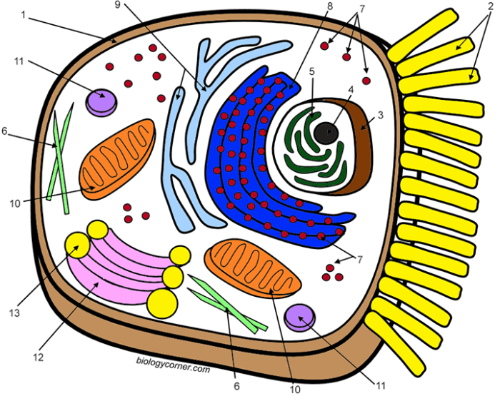
Animal cell coloring key biology corner – Animal cells are the fundamental building blocks of animals, exhibiting a complex internal organization crucial for their diverse functions. Understanding their structure is key to grasping the processes of life itself. This section will explore the major components of an animal cell and their roles, highlighting key differences from plant cells.The animal cell is characterized by a variety of membrane-bound organelles, each performing specialized tasks that contribute to the overall functioning of the cell.
These organelles work in a coordinated manner to maintain homeostasis and carry out essential cellular processes such as metabolism, growth, and reproduction. Unlike plant cells, animal cells lack a rigid cell wall and chloroplasts, features that significantly influence their structure and function.
Major Organelles and Their Functions
The internal structure of an animal cell is incredibly intricate. Several key organelles contribute to its overall function. These organelles, working in concert, ensure the cell’s survival and proper operation.
- Nucleus: The control center of the cell, containing the genetic material (DNA) which directs cellular activities.
- Ribosomes: Sites of protein synthesis, crucial for building the proteins necessary for cell structure and function. They can be free-floating in the cytoplasm or attached to the endoplasmic reticulum.
- Endoplasmic Reticulum (ER): A network of membranes involved in protein and lipid synthesis and transport. The rough ER (studded with ribosomes) synthesizes proteins, while the smooth ER synthesizes lipids and detoxifies substances.
- Golgi Apparatus (Golgi Body): Modifies, sorts, and packages proteins and lipids for secretion or delivery to other organelles.
- Mitochondria: The “powerhouses” of the cell, generating energy (ATP) through cellular respiration.
- Lysosomes: Membrane-bound sacs containing digestive enzymes that break down waste materials and cellular debris.
- Cytoskeleton: A network of protein filaments that provides structural support and facilitates cell movement.
- Centrioles: Play a crucial role in cell division, organizing microtubules during mitosis and meiosis.
Differences Between Plant and Animal Cells
While both plant and animal cells are eukaryotic (possessing a membrane-bound nucleus), several key distinctions exist. These differences reflect the contrasting lifestyles and functions of plants and animals.
| Feature | Plant Cell | Animal Cell |
|---|---|---|
| Cell Wall | Present (rigid, made of cellulose) | Absent |
| Chloroplasts | Present (sites of photosynthesis) | Absent |
| Vacuoles | Large central vacuole (stores water and nutrients) | Small or absent |
| Shape | Typically rectangular or polygonal | Variable, often round |
| Cell Size | Generally larger | Generally smaller |
Cell Membrane Structure and Function
The cell membrane, also known as the plasma membrane, is a selectively permeable barrier that encloses the cytoplasm and regulates the passage of substances into and out of the cell. This crucial membrane maintains cellular integrity and controls the internal environment.The cell membrane is primarily composed of a phospholipid bilayer, a double layer of phospholipid molecules arranged with their hydrophilic (water-loving) heads facing outward and their hydrophobic (water-fearing) tails facing inward.
Embedded within this bilayer are various proteins that perform diverse functions, including transport, cell signaling, and cell adhesion. This fluid mosaic model describes the dynamic nature of the membrane, where components are constantly moving and interacting. The selective permeability of the membrane ensures that essential nutrients enter the cell while waste products and harmful substances are kept out.
This regulation is vital for maintaining cellular homeostasis and preventing damage. The process involves various mechanisms, including simple diffusion, facilitated diffusion, active transport, and endocytosis/exocytosis. For instance, glucose enters the cell via facilitated diffusion using specific glucose transporter proteins, while larger molecules may enter via endocytosis.
Coloring and Identifying Animal Cell Organelles: Animal Cell Coloring Key Biology Corner

This section will guide you through the process of coloring and identifying the various organelles within an animal cell. Understanding the function and appearance of each organelle is crucial for comprehending the overall workings of the cell. We will provide a table to assist in your coloring activity, followed by a step-by-step guide.
Accurate representation of cell organelles is important for visualizing their functions and relationships within the cell. However, depicting three-dimensional structures on a two-dimensional surface presents certain challenges, which will be discussed later.
Animal Cell Organelle Chart, Animal cell coloring key biology corner
The following table provides a list of common animal cell organelles, their functions, suggested coloring codes, and distinguishing features to aid in your coloring exercise. Remember, these are suggestions; feel free to adapt the colors to your preference, ensuring clarity and distinction between organelles.
| Organelle Name | Function | Color Code | Distinguishing Features |
|---|---|---|---|
| Cell Membrane | Regulates what enters and leaves the cell. | Light Blue | Outer boundary; thin, continuous layer. |
| Cytoplasm | Gel-like substance filling the cell; location of many metabolic processes. | Light Yellow | Fills the space between organelles. |
| Nucleus | Contains genetic material (DNA); controls cell activities. | Dark Purple | Large, usually centrally located; often contains a visible nucleolus. |
| Nucleolus | Produces ribosomes. | Darker Purple | Smaller, dense structure within the nucleus. |
| Ribosomes | Synthesize proteins. | Dark Grey/Black | Small dots scattered throughout the cytoplasm and on the rough endoplasmic reticulum. |
| Rough Endoplasmic Reticulum (RER) | Protein synthesis and modification; studded with ribosomes. | Medium Blue | Network of interconnected membranes; appears rough due to ribosomes. |
| Smooth Endoplasmic Reticulum (SER) | Lipid synthesis and detoxification. | Light Green | Network of interconnected membranes; lacks ribosomes, appearing smooth. |
| Golgi Apparatus (Golgi Body) | Processes and packages proteins and lipids. | Light Orange | Stack of flattened sacs; often located near the RER. |
| Mitochondria | Produce energy (ATP) through cellular respiration. | Red | Bean-shaped or sausage-shaped organelles; often depicted with inner folds (cristae). |
| Lysosomes | Break down waste materials and cellular debris. | Dark Green | Small, membrane-bound sacs; often depicted as irregularly shaped. |
| Centrioles | Involved in cell division. | Pink | Paired cylindrical structures usually located near the nucleus. |
Step-by-Step Coloring Guide
This guide Artikels the steps for coloring a typical animal cell diagram, using the color codes suggested in the table above. Remember to use a light hand for shading to avoid obscuring the details of the organelles.
- Begin by coloring the cytoplasm with light yellow, ensuring to fill the entire area within the cell membrane.
- Next, color the nucleus dark purple and the nucleolus a slightly darker shade of purple.
- Color the cell membrane light blue, ensuring a clear distinction from the cytoplasm.
- Add the mitochondria in red, representing their bean-like shape.
- Color the rough endoplasmic reticulum medium blue, remembering to depict the ribosomes (dark grey/black) attached to its surface.
- Color the smooth endoplasmic reticulum light green, highlighting its smooth appearance compared to the RER.
- Add the Golgi apparatus in light orange, showing its characteristic stacked structure.
- Represent the lysosomes with dark green, remembering their irregular shape.
- Finally, add the centrioles in pink, near the nucleus.
Challenges of Representing Three-Dimensional Structures in Two Dimensions
Accurately depicting the three-dimensional nature of organelles, such as the intricate folding within the mitochondria (cristae) or the interconnected network of the endoplasmic reticulum, presents a significant challenge in a two-dimensional coloring activity. The limitations of flat surfaces necessitate simplification and symbolic representation. For example, the cristae within mitochondria are often shown as simple lines or shading rather than their complex three-dimensional folds.
Similarly, the extensive network of the endoplasmic reticulum is often represented as a simplified diagrammatic structure, sacrificing the full complexity of its interconnectedness.
Illustrating Animal Cell Components

This section delves into the detailed structure and function of key animal cell components, providing descriptions suitable for illustrative purposes. Understanding these structures is crucial for comprehending the overall function of the cell. We will focus on the nucleus, mitochondria, and the endoplasmic reticulum-Golgi apparatus system.
The Nucleus: Structure and Components
The nucleus, the cell’s control center, is a membrane-bound organelle housing the cell’s genetic material. Its structure is complex and integral to cellular function. The nuclear envelope, a double membrane punctuated by nuclear pores, regulates the passage of molecules between the nucleus and the cytoplasm. Within the nucleus, the nucleolus is a dense region responsible for ribosome biogenesis – the creation of ribosomes, crucial for protein synthesis.
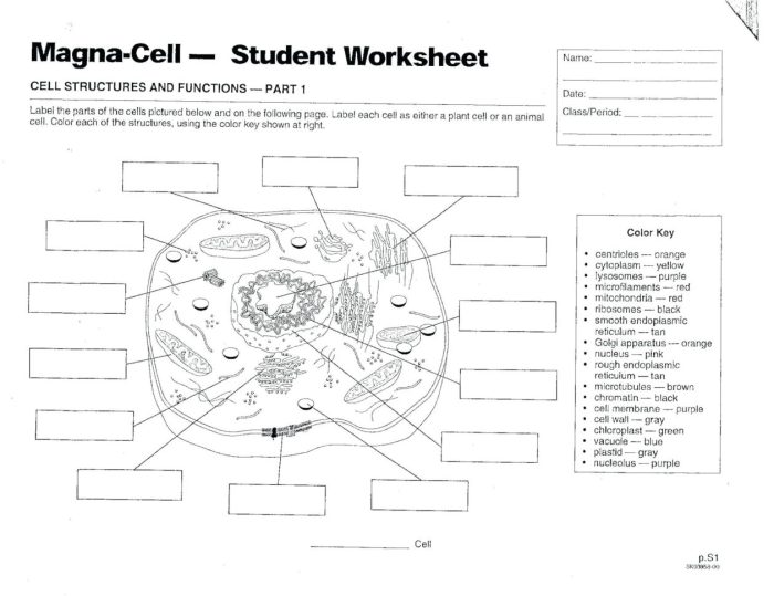
Understanding animal cell structures is simplified using the animal cell coloring key from Biology Corner; it’s a great starting point for learning. To further practice visualizing these structures, you might find a helpful resource in this animal blank coloring sheet , allowing you to draw and label the organelles yourself. Returning to the Biology Corner key, remember to check your work against the accurate depictions provided.
Chromatin, a complex of DNA and proteins, condenses into chromosomes during cell division, carrying the genetic instructions for the cell. An illustration would show the nuclear envelope as a double line with pores depicted as small openings, the nucleolus as a darker, round structure within the nucleus, and the chromatin as a diffuse, thread-like network filling the nuclear space.
Mitochondrial Cellular Respiration
Mitochondria are often referred to as the “powerhouses” of the cell because they are the sites of cellular respiration. This process converts chemical energy from nutrients into a usable form of energy, ATP (adenosine triphosphate). Cellular respiration can be illustrated as a multi-stage process. First, glycolysis breaks down glucose in the cytoplasm, yielding a small amount of ATP and pyruvate.
Pyruvate then enters the mitochondria, where it undergoes the Krebs cycle (citric acid cycle) in the mitochondrial matrix. This cycle generates more ATP, NADH, and FADH2. Finally, oxidative phosphorylation, occurring across the inner mitochondrial membrane, utilizes the electron carriers NADH and FADH2 to drive ATP synthesis through chemiosmosis. The illustration would depict the mitochondrion as a bean-shaped organelle with a folded inner membrane (cristae) and a matrix, highlighting the different locations of glycolysis, the Krebs cycle, and oxidative phosphorylation.
The final product, ATP, should be prominently featured.
Endoplasmic Reticulum and Golgi Apparatus: Protein Modification and Transport
The endoplasmic reticulum (ER) and Golgi apparatus work together in a coordinated manner to modify and transport proteins. The rough ER, studded with ribosomes, synthesizes proteins destined for secretion or membrane incorporation. These proteins then move to the smooth ER, which is involved in lipid synthesis and detoxification. From the ER, proteins are transported to the Golgi apparatus, a series of flattened sacs (cisternae).
Here, proteins undergo further processing, including glycosylation (addition of sugar molecules) and folding, before being packaged into vesicles for transport to their final destinations – either within the cell or for secretion outside the cell. An illustration would show the rough ER as a network of interconnected membranes with ribosomes attached, the smooth ER as a similar network but without ribosomes, and the Golgi apparatus as a stack of flattened sacs with vesicles budding off.
Arrows could illustrate the flow of proteins from the ribosomes to the ER, then to the Golgi, and finally to their destinations.
Practical Applications of Animal Cell Knowledge
Understanding animal cell structure and function is not merely an academic exercise; it forms the bedrock of numerous crucial advancements in medicine and biotechnology. This knowledge translates directly into tangible improvements in human health and well-being, impacting diagnosis, treatment, and the development of new therapies.Our comprehension of animal cell processes is fundamental to medical breakthroughs. The intricate mechanisms within cells, from DNA replication to protein synthesis, are targets for therapeutic intervention.
Disruptions in these processes often underlie diseases, and manipulating these pathways can lead to effective treatments.
Animal Cell Research in Medicine
The study of animal cells is paramount in various medical research areas. For instance, understanding the mechanisms of cell division is crucial for cancer research. Cancer is characterized by uncontrolled cell growth, and investigating the cellular processes involved allows scientists to develop targeted therapies that selectively inhibit tumor growth while minimizing damage to healthy cells. Similarly, research into cellular signaling pathways is essential for understanding and treating autoimmune diseases, where the body’s immune system mistakenly attacks its own cells.
Studies on cellular responses to pathogens are critical for developing vaccines and antiviral treatments. In neuroscience, research on neuronal cells helps in understanding neurological disorders and developing new therapies for conditions like Alzheimer’s and Parkinson’s disease.
Developing New Medicines and Treatments
Knowledge of animal cell function directly informs the development of new medicines and treatments. Drug discovery relies heavily on understanding how drugs interact with cellular components and pathways. For example, many drugs target specific receptors or enzymes within cells to modulate their activity. This requires detailed knowledge of the structure and function of these cellular targets. Furthermore, animal cell cultures are widely used in drug testing to assess the efficacy and safety of new compounds before human trials.
This process helps identify potential side effects and optimize drug dosage. Advances in gene editing technologies, such as CRISPR-Cas9, rely on a thorough understanding of cellular mechanisms to precisely modify genes within animal cells, offering potential cures for genetic diseases.
Ethical Considerations in Animal Cell Research
While research involving animal cells offers immense potential for medical advancements, ethical considerations are paramount. The use of animal-derived cells raises questions about animal welfare and the potential for suffering. Researchers must adhere to strict ethical guidelines, ensuring that animal cells are obtained and used responsibly, minimizing any potential harm to animals. Furthermore, the use of human cells and tissues raises questions about informed consent and data privacy.
Strict regulations and ethical review boards are essential to oversee research involving animal and human cells, balancing the potential benefits with the ethical implications. Open and transparent discussions regarding the ethical implications of animal cell research are vital to ensuring responsible scientific progress.
FAQs
What are some common mistakes students make when coloring animal cells?
Common mistakes include inaccurate coloring of organelles based on size and location, inconsistent color usage, and neglecting to label the organelles.
Are there any online resources besides Biology Corner for further learning?
Yes, many websites and educational platforms offer interactive simulations, 3D models, and virtual labs focusing on animal cell structure and function. A quick search for “interactive animal cell model” will yield numerous results.
How can I make the coloring activity more engaging for younger learners?
Incorporate creative elements like adding labels with fun fonts, using different textures, or turning the completed coloring into a small poster or presentation.
What are the ethical considerations surrounding animal cell research?
Ethical considerations revolve around the source of animal cells (e.g., ensuring humane treatment of animals if cells are derived from them) and responsible use of research findings, avoiding misuse or unethical applications.


