Understanding Animal Cell Structure
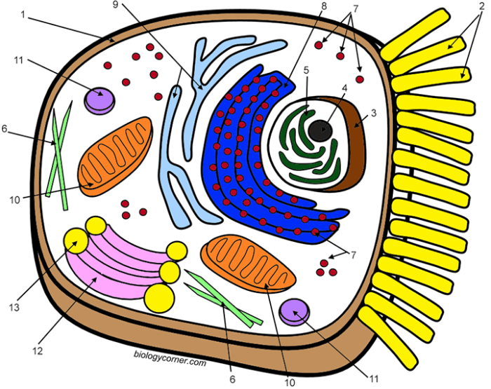
Animal cell coloring answers key – Animal cells are the fundamental building blocks of animals, exhibiting a complex internal organization crucial for their survival and function. Understanding their structure provides insight into the intricate processes occurring within these microscopic units. This section details the key components of animal cells and their roles.
Major Organelles and Their Functions
Animal cells contain various organelles, each with a specific function contributing to the cell’s overall operation. These organelles work in a coordinated manner to maintain homeostasis and carry out essential life processes.
| Organelle | Function | Description | Illustration |
|---|---|---|---|
| Nucleus | Houses genetic material (DNA), controls cell activities. | A large, membrane-bound organelle containing chromosomes. It’s the control center of the cell, dictating protein synthesis and cell division. | A large, roughly spherical structure with a darker area (nucleolus) visible within. The outer membrane is continuous with the endoplasmic reticulum. |
| Ribosomes | Protein synthesis. | Small, granular structures found free in the cytoplasm or attached to the endoplasmic reticulum. They are the sites where amino acids are linked together to form proteins. | Tiny dots, either scattered throughout the cytoplasm or clustered on the surface of the endoplasmic reticulum. |
| Endoplasmic Reticulum (ER) | Protein and lipid synthesis, transport. | A network of interconnected membranes forming channels throughout the cytoplasm. Rough ER (with ribosomes) synthesizes proteins, while smooth ER synthesizes lipids and detoxifies substances. | A network of interconnected flattened sacs and tubules extending throughout the cytoplasm. Rough ER appears studded with ribosomes. |
| Golgi Apparatus (Golgi Body) | Modifies, sorts, and packages proteins and lipids. | A stack of flattened, membrane-bound sacs. It receives proteins and lipids from the ER, modifies them, and packages them into vesicles for transport. | A stack of flattened sacs, resembling a stack of pancakes, with vesicles budding off from the edges. |
| Mitochondria | Cellular respiration, ATP production. | Rod-shaped or oval organelles with a double membrane. They are the “powerhouses” of the cell, generating energy in the form of ATP. | Bean-shaped organelles with a folded inner membrane (cristae). |
| Lysosomes | Waste breakdown, digestion. | Membrane-bound sacs containing digestive enzymes. They break down waste materials, cellular debris, and foreign substances. | Small, membrane-bound sacs containing dark, granular material. |
| Centrioles | Cell division. | Paired cylindrical structures involved in organizing microtubules during cell division. | Two short, cylindrical structures positioned perpendicular to each other near the nucleus. |
| Cytoplasm | Supports organelles, site of many metabolic reactions. | The jelly-like substance filling the cell, containing the organelles and cytoskeleton. | The clear, gel-like substance filling the cell, surrounding the organelles. |
| Cell Membrane | Regulates passage of substances into and out of the cell. | A thin, flexible outer boundary of the cell. It is selectively permeable, controlling what enters and exits the cell. | A thin, continuous line surrounding the cell, representing the boundary between the inside and outside environments. |
Plant and Animal Cell Differences
While both plant and animal cells are eukaryotic, meaning they have a membrane-bound nucleus, they differ significantly in structure. Plant cells possess features absent in animal cells, reflecting their different functions and lifestyles. A key difference is the presence of a cell wall and chloroplasts in plant cells.
Cell Membrane and Its Role in Cell Function, Animal cell coloring answers key
The cell membrane, also known as the plasma membrane, is a selectively permeable barrier that regulates the movement of substances into and out of the cell. This is crucial for maintaining the cell’s internal environment and carrying out essential metabolic processes. The membrane’s structure, primarily a phospholipid bilayer with embedded proteins, allows for controlled transport mechanisms, including diffusion, osmosis, and active transport.
This controlled exchange ensures the cell receives necessary nutrients and eliminates waste products. The membrane also plays a vital role in cell signaling and communication.
Coloring Activities and their Educational Value
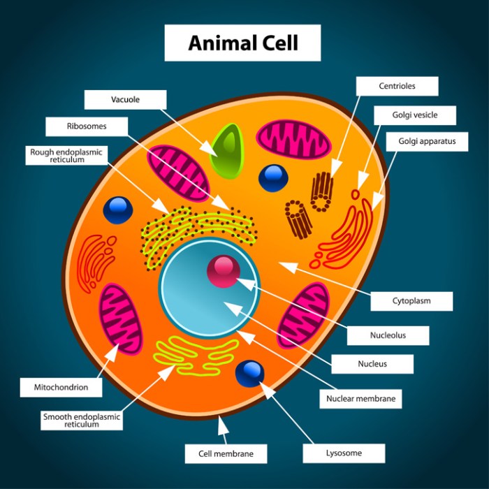
Coloring activities offer a surprisingly effective and engaging method for learning about complex biological structures like the animal cell. This hands-on approach transforms the often-abstract concepts of cellular biology into a tangible and memorable experience, significantly enhancing comprehension and retention. The act of coloring, coupled with labeling organelles, actively involves the learner, fostering a deeper understanding than passive learning methods alone.Coloring improves memory and understanding of complex biological concepts by engaging multiple learning styles simultaneously.
Visual learners benefit directly from the pictorial representation of the cell and its components. Kinesthetic learners engage through the physical act of coloring and labeling. Furthermore, the process of associating specific colors with specific organelles strengthens memory recall. The repetitive nature of coloring and labeling reinforces learning, making the information more readily accessible for later retrieval.
This multi-sensory approach proves particularly beneficial for students who struggle with abstract concepts, offering an alternative pathway to understanding.
Comparison of Coloring Methods for Teaching Animal Cell Structure
Different coloring methods cater to varying learning preferences and educational goals. A simple black and white worksheet allows students to use their creativity in choosing colors, fostering individual expression while still focusing on accurate organelle placement and labeling. Conversely, a pre-colored worksheet with labeled organelles can serve as a visual reference for students to copy, providing a structured approach for those who benefit from clear examples.
A more advanced method might involve providing students with a blank worksheet and a list of organelles and their functions, requiring them to research and color accordingly, encouraging independent learning and critical thinking. Each method presents unique advantages, making it crucial to select the approach best suited to the students’ needs and learning objectives.
Example Coloring Worksheet with Detailed Labels
Imagine a worksheet depicting a typical animal cell, roughly circular in shape. The cell membrane, a thin outer boundary, should be colored a light blue, labeled clearly as “Cell Membrane.” The cytoplasm, the jelly-like substance filling the cell, could be a pale yellow, labeled “Cytoplasm.” The nucleus, a large, centrally located organelle, might be colored dark purple and labeled “Nucleus.” Within the nucleus, a smaller, darker area representing the nucleolus could be labeled “Nucleolus.” The rough endoplasmic reticulum (RER), studded with ribosomes, could be depicted as a network of interconnected, slightly darker blue tubes, labeled “Rough Endoplasmic Reticulum (RER).” The smooth endoplasmic reticulum (SER), lacking ribosomes, could be a lighter shade of blue, labeled “Smooth Endoplasmic Reticulum (SER).” The Golgi apparatus, a stack of flattened sacs, could be colored light green and labeled “Golgi Apparatus.” Mitochondria, the powerhouses of the cell, could be depicted as bean-shaped structures in a reddish-brown, labeled “Mitochondria.” Lysosomes, small, spherical organelles, could be colored dark orange, labeled “Lysosomes.” Finally, ribosomes, tiny dots scattered throughout the cytoplasm and on the RER, could be depicted as small dark purple dots, labeled “Ribosomes.” This detailed labeling and coloring exercise provides a comprehensive visual representation of the animal cell’s key components.
Advanced Applications of Animal Cell Knowledge
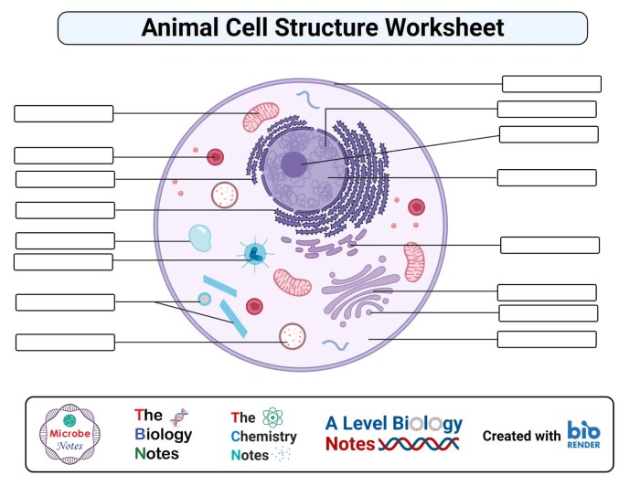
Understanding animal cell structure is fundamental to comprehending a wide range of biological processes and their implications for health and disease. A deep understanding of the cell’s intricate machinery allows us to connect its components to overall cellular function and, ultimately, to the functioning of the organism as a whole.
Animal Cell Structure and Cellular Processes
The structure of an animal cell directly influences its ability to perform vital cellular processes. For example, the mitochondria, with their folded inner membranes (cristae), provide a large surface area for the electron transport chain, crucial for cellular respiration and ATP production. The efficient production of ATP fuels numerous cellular activities, from muscle contraction to protein synthesis. Similarly, the endoplasmic reticulum (ER) and ribosomes work in concert for protein synthesis.
Ribosomes, either free-floating in the cytoplasm or bound to the rough ER, translate mRNA into polypeptide chains. The ER then processes and folds these proteins, ensuring proper functionality. Disruptions in either the mitochondria or the ER-ribosome system can severely impair cellular function.
Animal Cells in the Human Body
Animal cells are the building blocks of all human tissues and organs. Epithelial cells form protective layers in the skin and lining of internal organs. Muscle cells, with their specialized contractile proteins, enable movement. Neurons, with their long axons and dendrites, facilitate communication throughout the nervous system. Understanding the unique structures of these cells helps explain their diverse functions.
For instance, the elongated shape of neurons allows for efficient signal transmission over long distances, while the tightly packed nature of epithelial cells creates a barrier against pathogens. Different cell types exhibit variations in organelle composition and abundance, reflecting their specialized roles. For example, muscle cells contain numerous mitochondria to support their high energy demands, while secretory cells have a well-developed Golgi apparatus for packaging and secretion of proteins.
Disruptions in Animal Cell Structure and Disease
Many diseases arise from disruptions in animal cell structure or function. For example, cystic fibrosis results from a defect in a protein that regulates chloride ion transport across cell membranes, leading to thick mucus buildup in the lungs and other organs. Cancer is characterized by uncontrolled cell growth and division, often stemming from mutations affecting cell cycle regulation or DNA repair mechanisms.
Mitochondrial diseases, resulting from defects in mitochondrial DNA or proteins, can manifest in a wide range of symptoms affecting energy production in various tissues. These examples highlight the critical link between cellular structure, function, and overall health.
Microscopy Techniques for Visualizing Animal Cells
Various microscopy techniques are employed to visualize animal cells and their organelles. Light microscopy provides a general overview of cell structure and morphology, while electron microscopy offers much higher resolution, allowing for visualization of individual organelles and even macromolecular structures. Transmission electron microscopy (TEM) provides detailed internal structures, while scanning electron microscopy (SEM) reveals surface details. Fluorescence microscopy allows for the visualization of specific proteins or organelles using fluorescently labeled antibodies or probes.
These techniques are crucial for understanding cell biology and diagnosing diseases. For example, using immunofluorescence microscopy, researchers can identify specific proteins associated with cancer cells, contributing to improved diagnosis and treatment strategies.
Finding the answers for your animal cell coloring sheet can be challenging, requiring careful observation of organelles. A fun break from the scientific detail might be found with a creative activity like the animal cartwheel coloring sheet , offering a different type of visual engagement. Afterwards, returning to the precision needed for the animal cell coloring answers key will be easier after this refreshing change of pace.
Creating Visual Aids for Animal Cell Structure
Visual aids are crucial for understanding complex biological processes like those occurring within an animal cell. Effective visuals can transform abstract concepts into concrete, easily grasped representations, facilitating better comprehension and retention of information. The following sections detail the creation of several visual aids to illustrate key aspects of animal cell structure and function.
Protein Synthesis Flowchart
This flowchart depicts the journey of a protein from its genetic blueprint to its final destination within the cell. It begins in the nucleus with DNA transcription, where the genetic code is copied into messenger RNA (mRNA). The mRNA then moves to the ribosomes, either free-floating in the cytoplasm or bound to the endoplasmic reticulum (ER). At the ribosomes, translation occurs – the mRNA code is read, and amino acids are assembled into a polypeptide chain.
This chain folds into a specific three-dimensional structure, often with assistance from chaperone proteins. Finally, the completed protein is transported to its designated location within or outside the cell, perhaps via the Golgi apparatus for modification and packaging. The flowchart would visually represent these steps with boxes and arrows, clearly labeling each stage and the organelles involved.
Three-Dimensional Model of an Animal Cell
An accurate 3D model of an animal cell would be roughly spherical, though the exact shape can vary depending on the cell type and its environment. A typical size would range from 10 to 30 micrometers in diameter. The nucleus, a large, centrally located organelle, would be clearly visible, representing the cell’s control center. The endoplasmic reticulum (ER), a network of interconnected membranes, would be depicted extending throughout the cytoplasm.
Ribosomes, small granular structures, would be shown either attached to the ER or free in the cytoplasm. The Golgi apparatus, a stack of flattened sacs, would be positioned near the ER, indicating its role in protein modification and transport. Mitochondria, the powerhouses of the cell, would be scattered throughout, illustrating their energy-producing function. Lysosomes, small vesicles containing digestive enzymes, would be visible, and the cell membrane, a thin outer boundary, would enclose the entire structure.
The relative sizes and positions of these organelles would be accurately represented to reflect their actual proportions and spatial relationships within the cell.
Cell Membrane Diagram
This diagram would illustrate the fluid mosaic model of the cell membrane, highlighting its dynamic nature. The phospholipid bilayer, the membrane’s core structure, would be shown with hydrophilic heads facing outward and hydrophobic tails inward. Embedded within this bilayer would be various proteins, including integral proteins extending across the membrane and peripheral proteins attached to its surface. These proteins would be depicted performing different functions, such as transport, cell signaling, and cell adhesion.
The diagram would also show cholesterol molecules interspersed within the phospholipid bilayer, contributing to membrane fluidity and stability. The extracellular matrix, a network of molecules outside the cell membrane, would be depicted interacting with the membrane proteins, facilitating cell-to-cell communication and support. The diagram would clearly show the selective permeability of the membrane, illustrating how certain substances can pass through while others are blocked.
Animal Cell Division Illustration
This illustration would depict the process of mitosis, the type of cell division that produces two identical daughter cells from a single parent cell. The stages of mitosis – prophase, metaphase, anaphase, and telophase – would be shown sequentially. Prophase would depict the condensation of chromosomes, the breakdown of the nuclear envelope, and the formation of the mitotic spindle.
Metaphase would show the chromosomes aligned at the metaphase plate. Anaphase would illustrate the separation of sister chromatids and their movement towards opposite poles of the cell. Telophase would show the reformation of the nuclear envelope and the division of the cytoplasm (cytokinesis), resulting in two genetically identical daughter cells. The illustration would clearly show the changes in chromosome structure and the role of the mitotic spindle in ensuring accurate chromosome segregation.
The differences between animal and plant cell division (e.g., the formation of a cleavage furrow in animal cells versus a cell plate in plant cells) could also be highlighted.
Popular Questions: Animal Cell Coloring Answers Key
What are some good resources for printable animal cell coloring pages?
Many educational websites and online resources offer free printable animal cell coloring pages. A simple web search should yield numerous results.
How can I make my own animal cell coloring worksheet?
Use a drawing program or even hand-draw a simple animal cell diagram. Label the organelles clearly and provide space for students to color them in.
Are there any apps or online tools that can help with animal cell coloring and learning?
Several educational apps and interactive websites offer virtual cell models and coloring activities. A quick search in your app store or online will provide various options.


