Introduction to Animal Cell Structure: Animal Cell Coloring Answers Key

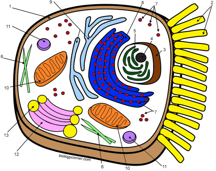
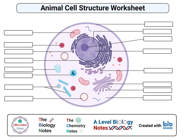
Animal cell coloring answers key – Yo, Medan peeps! Let’s dive into the amazing world of animal cells. Think of them as the tiny building blocks that make up you, me, and every other animal out there. These cells are super complex, packed with different parts that all work together like a well-oiled machine. Understanding their structure is key to understanding how life itself works.Animal cells, unlike plant cells, don’t have cell walls or chloroplasts.
They’re more flexible and diverse in shape, adapting to their various roles in the body. Let’s explore some of the key players within these microscopic powerhouses.
The Nucleus: The Cell’s Brain
The nucleus is the control center of the animal cell, kinda like the brain of the operation. It’s enclosed by a double membrane called the nuclear envelope, which has tiny pores that regulate what goes in and out. Inside, you’ll find the cell’s genetic material, DNA, organized into chromosomes. DNA holds the blueprints for building and running the entire cell.
Think of it as the ultimate instruction manual. The nucleolus, a dense region within the nucleus, is responsible for making ribosomes—the protein factories of the cell.
Cytoplasm: The Cell’s Busy Hub
The cytoplasm is the jelly-like substance filling the cell, excluding the nucleus. It’s a bustling environment where many cellular processes occur. It’s not just empty space; it’s a dynamic mixture of water, salts, and various organelles. The cytoskeleton, a network of protein fibers, provides structural support and helps with cell movement and transport within the cytoplasm. It’s like the cell’s internal highway system.
Cell Membrane: The Gatekeeper
The cell membrane is the outer boundary of the animal cell, a selectively permeable barrier. This means it controls what enters and exits the cell, maintaining a stable internal environment. It’s made up of a lipid bilayer with embedded proteins that act as channels and pumps for transporting molecules. Imagine it as a bouncer at a club, carefully selecting who gets in and who stays out.
Mitochondria: The Powerhouses
Mitochondria are often called the “powerhouses” of the cell because they generate most of the cell’s energy in the form of ATP (adenosine triphosphate). They have their own DNA and ribosomes, suggesting an endosymbiotic origin – they were once independent organisms. These little energy factories are crucial for cellular respiration, the process that converts nutrients into usable energy.
They’re like tiny power plants within the cell.
Ribosomes: The Protein Factories, Animal cell coloring answers key
Ribosomes are the protein synthesis machines of the cell. They’re responsible for translating the genetic code from DNA into proteins, the workhorses of the cell. Ribosomes can be found free-floating in the cytoplasm or attached to the endoplasmic reticulum (ER). Think of them as the construction workers, building all the proteins needed for the cell’s functions.
Major Organelles and Their Functions
| Organelle Name | Function | Visual Description |
|---|---|---|
| Nucleus | Houses DNA, controls cell activities | A large, round structure usually near the center of the cell. |
| Cytoplasm | Jelly-like substance, site of many cellular processes | Fills the cell, surrounds organelles. |
| Cell Membrane | Regulates what enters and exits the cell | Thin, flexible outer boundary of the cell. |
| Mitochondria | Generates ATP (cellular energy) | Rod-shaped or oval structures with a folded inner membrane. |
| Ribosomes | Synthesizes proteins | Small, granular structures, found free or attached to ER. |
Analyzing the Coloring Process

Coloring an animal cell isn’t just a fun activity; it’s a sneaky way to boost your brainpower, Medan style! It’s like leveling up your understanding of biology without even realizing it. Think of it as a supercharged study session disguised as a creative project.This activity cleverly taps into several cognitive skills. By carefully following the instructions and matching colors to specific organelles, you’re actively engaging in problem-solving and enhancing your attention to detail.
It’s a low-key exercise in visual processing, memory recall, and hand-eye coordination. Plus, the act of coloring itself can be incredibly calming and meditative, improving focus and concentration.
Spatial Relationships and Organelle Sizes
Accurate coloring helps you visualize the spatial arrangement of organelles within the cell. For example, you’ll see how the nucleus, usually a large, centrally located structure, interacts with the surrounding cytoplasm, which fills the entire cell. Similarly, you’ll visually grasp the relative sizes of organelles like mitochondria (relatively large, sausage-shaped) versus ribosomes (tiny dots scattered throughout the cytoplasm).
This visual representation significantly enhances your understanding of the cell’s three-dimensional structure, moving beyond a simple two-dimensional diagram. Imagine trying to explain the arrangement of furniture in a room; a drawing helps, but actually arranging the furniture gives you a far better understanding of the space. Similarly, coloring an animal cell solidifies your understanding of its internal organization.
Connection Between Accurate Coloring and Cellular Function
The act of correctly identifying and coloring each organelle directly correlates with understanding its function. For instance, accurately coloring the mitochondria a vibrant color helps you remember its role as the powerhouse of the cell, generating energy through cellular respiration. Similarly, coloring the Golgi apparatus, a flattened stack of membranes, reinforces its function in processing and packaging proteins. This visual association creates a stronger neural connection between the visual representation and the functional role of each organelle, leading to a more profound and lasting understanding of the complex processes within an animal cell.
It’s like learning vocabulary – seeing the word written and associating it with its meaning creates a stronger memory trace than just hearing it.
Understanding the intricacies of an animal cell can be challenging, but a visual approach can help. The detailed diagrams found in “animal cell coloring answers key” worksheets provide a great starting point. For a fun break, consider supplementing your studies with some engaging visuals, like those available at coloring pages of animals , which can help reinforce basic animal anatomy.
Returning to the “animal cell coloring answers key,” remember that accurate coloring helps solidify your understanding of cell structures and their functions.


