Analyzing Animal Cell Structure for the Coloring Page
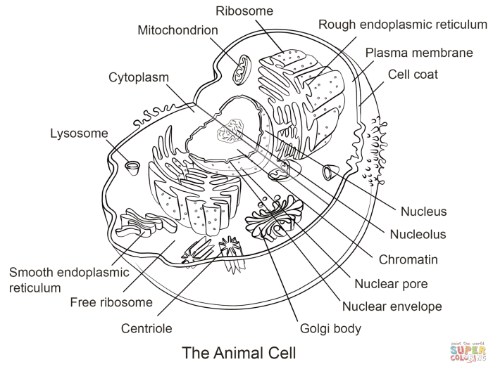
Animal cell coloring page 77 – This section will guide you through the key components of an animal cell, providing descriptions and visual details perfect for your coloring page. Understanding the structure and function of each organelle will bring your artwork to life and enhance your understanding of cellular biology. We will focus on the major organelles, providing sufficient detail for a vibrant and informative coloring page.
Cell Membrane
The cell membrane is the outer boundary of the animal cell. Imagine it as a flexible, protective skin. For your coloring page, depict it as a thin, continuous line surrounding the entire cell. It’s selectively permeable, meaning it controls what enters and exits the cell. You could color it a light blue to represent its fluid nature.
Think of it as a gatekeeper, allowing essential nutrients in and waste products out.
Cytoplasm
The cytoplasm is the jelly-like substance filling the cell. It’s the internal environment where many cellular processes occur. On your coloring page, represent it as a light, even shading within the cell membrane, but outside the nucleus and other organelles. You might color it a pale yellow or light green to suggest its fluid consistency. Many cellular reactions take place within this gel-like substance.
Nucleus
The nucleus is the control center of the cell, containing the cell’s genetic material (DNA). Picture it as a large, round structure within the cell. For your coloring page, draw it as a large circle near the center of the cell and color it a darker shade, perhaps a light purple, to distinguish it from the cytoplasm. You can even add a small, darker circle within the nucleus to represent the nucleolus, where ribosomes are made.
Mitochondria
Mitochondria are the powerhouses of the cell, generating energy (ATP) through cellular respiration. Imagine them as bean-shaped structures scattered throughout the cytoplasm. On your coloring page, draw several small, bean-shaped figures throughout the cytoplasm. Color them a reddish-brown to reflect their energy-producing role.
Ribosomes
Ribosomes are tiny structures responsible for protein synthesis. They are too small to be individually detailed on a coloring page, so you can represent them as small dots scattered throughout the cytoplasm, perhaps slightly clustered around the endoplasmic reticulum (if you choose to include it). Color them a dark grey or black to represent their dense structure.
Golgi Apparatus
The Golgi apparatus is a stack of flattened sacs that modifies, sorts, and packages proteins. Visualize it as a series of stacked pancakes or flattened sacs near the nucleus. For your coloring page, draw a series of slightly curved, parallel lines near the nucleus and color them a light orange or tan. This structure is vital for the transport of proteins throughout the cell.
Simplified Diagram of an Animal Cell
Imagine a circle (the cell membrane) containing a large central circle (the nucleus – purple). Scattered within the cytoplasm (pale yellow) are bean-shaped structures (mitochondria – reddish-brown) and numerous small dots (ribosomes – dark grey). Near the nucleus, draw a stack of flattened sacs (Golgi apparatus – light orange). This simplified diagram clearly shows the key organelles and their relative positions within the cell.
Remember to label each organelle clearly.
Designing the Coloring Page
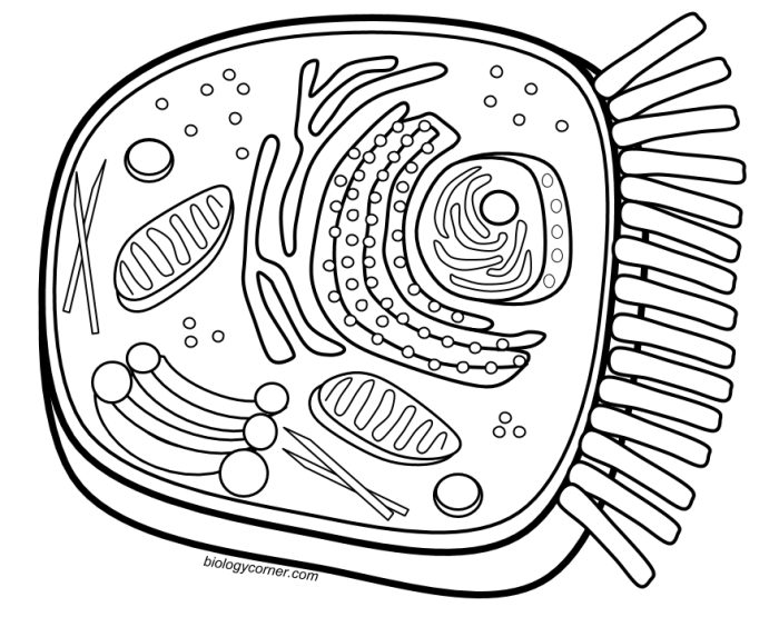
Creating engaging and informative coloring pages requires careful consideration of both visual appeal and educational accuracy. The goal is to make learning about the animal cell fun and accessible, catering to different skill levels. We will design two versions: a simplified version for younger learners and a more complex version for older children or those seeking a greater challenge.The design process involves choosing appropriate shapes and sizes for organelles, arranging them logically within the cell membrane, and ensuring clear labeling for easy identification.
The simplified version prioritizes clarity and ease of coloring, while the more complex version introduces greater detail and realism, encouraging a deeper understanding of the cell’s intricate structure.
Simplified Animal Cell Coloring Page Design
This version will utilize basic shapes to represent each organelle. The cell membrane will be a simple circle or oval. The nucleus will be a large, centrally located circle. The mitochondria will be depicted as smaller, bean-shaped ovals scattered throughout the cytoplasm. The endoplasmic reticulum will be represented by a network of simple lines, and the Golgi apparatus will be a stack of flattened sacs.
Lysosomes and ribosomes can be shown as small dots. Each organelle will be clearly labeled with simple, easy-to-read text. The overall color scheme should be bright and inviting, encouraging children to engage with the page. For example, the nucleus could be colored purple, the mitochondria orange, and the cytoplasm a light yellow.
Detailed Animal Cell Coloring Page Design
This version will incorporate more detailed representations of the organelles. The cell membrane will still be a circle or oval, but could include small details to represent the lipid bilayer. The nucleus will show a more realistic representation, perhaps including a nucleolus. Mitochondria will have more detailed inner structures depicted. The endoplasmic reticulum will be a more complex network of lines, differentiating between rough and smooth ER.
The Golgi apparatus will be shown with more precisely rendered cisternae. Lysosomes and ribosomes will still be shown as small dots but perhaps in greater numbers. The inclusion of additional organelles like the centrosome and vacuoles could also be considered. Labeling will be more detailed, providing both the name and a brief function for each organelle. This version allows for more creative coloring and a more in-depth understanding of the cell’s complexity.
For instance, the rough endoplasmic reticulum could be shaded darker to represent the ribosomes attached to it.
Animal cell coloring page 77 offers a detailed look at the inner workings of an animal cell. For a change of pace, and to explore animal anatomy in a different way, you might enjoy the fun illustrations found in animal alphabet coloring sheets. Returning to the cellular level, page 77 provides a great opportunity to learn about organelles and their functions within the animal cell.
Generating Alternative Representations: Animal Cell Coloring Page 77
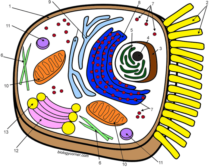
A coloring page is a valuable tool, but alternative visual representations can enhance understanding of animal cell structure. Different learning styles benefit from diverse approaches, making multiple representations crucial for comprehensive education. The following Artikels three alternatives, highlighting their advantages and disadvantages as educational tools.
Three-Dimensional Model Construction, Animal cell coloring page 77
Creating a three-dimensional model of an animal cell allows for a tangible and interactive learning experience. Students can use various materials like clay, styrofoam balls, pipe cleaners, and colored markers to represent different organelles. This hands-on approach fosters a deeper understanding of the spatial relationships between organelles and their relative sizes. The advantages include improved spatial reasoning and memory retention due to the tactile nature of the activity.
However, constructing accurate models can be time-consuming and require specialized materials. The accuracy of the representation is also dependent on the students’ understanding and skill in model-building. As an educational tool, this method excels in engaging kinesthetic learners and promoting collaborative learning within groups. The process of building the model reinforces the learning of organelle names and functions.
Animated Digital Representation
An animated digital representation, perhaps a short video or interactive simulation, can vividly illustrate the dynamic processes within an animal cell. This approach could showcase processes like protein synthesis, endocytosis, and exocytosis in a visually engaging way. The use of animation makes complex processes easier to understand.The benefits include the ability to show movement and change over time, something a static image cannot achieve.
However, creating high-quality animations requires specialized software and expertise, making it potentially resource-intensive. Moreover, the complexity of the animation could overwhelm students if not carefully designed. As an educational tool, this method excels in illustrating dynamic processes and engaging visual learners. Interactive simulations allow students to manipulate variables and explore the consequences, promoting active learning.
Detailed Diagram with Labeled Organelles
A detailed diagram, meticulously drawn and clearly labeled, provides a comprehensive overview of the animal cell’s structure. This method can incorporate realistic illustrations of organelles, highlighting their internal structures and functions. The diagram can be accompanied by a concise description of each organelle and its role within the cell.The advantage is its clarity and completeness; all key organelles are visible and labeled, providing a concise reference point.
However, a static diagram may lack the engagement of more dynamic representations. Furthermore, the effectiveness of this method depends on the quality of the diagram and the clarity of the labels and accompanying text. As an educational tool, this method is particularly effective for visual learners and provides a valuable reference for students to consult during learning.
It can be easily incorporated into textbooks, worksheets, and presentations.
FAQ Overview
What is the significance of the number “77” in “Animal Cell Coloring Page 77”?
The number “77” likely refers to a page number within a specific book, worksheet, or online resource. Without further context, its precise meaning remains unclear.
Are there different versions of animal cell coloring pages available?
Yes, various versions exist, ranging from simple diagrams for younger children to more complex representations for older students, incorporating more detail and labeling.
What materials are needed to create an animal cell coloring page?
You’ll need paper, coloring pencils or crayons, and potentially a printer to create a pre-designed template. Alternatively, you can draw the cell freehand.
How can I make my animal cell coloring page more engaging?
Incorporate vibrant colors, add interesting textures, and consider using different artistic styles to make the page more visually appealing and interactive.

