Introduction to Animal and Plant Cell Structures

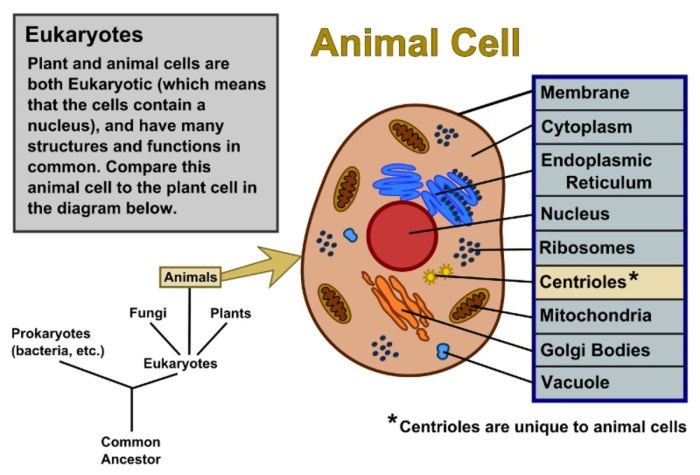
Animal and plant cell coloring pdf – Cells are the fundamental building blocks of all living organisms. Both animal and plant cells share some common features, but they also exhibit significant differences reflecting their distinct functions and roles in multicellular organisms. Understanding these similarities and differences is crucial to grasping the complexities of life.Animal and plant cells are both eukaryotic cells, meaning they possess a membrane-bound nucleus and other membrane-bound organelles.
However, several key structural components distinguish them. Plant cells are characterized by the presence of a rigid cell wall, chloroplasts, and a large central vacuole, features generally absent in animal cells. These structural differences directly impact the cells’ functions and overall organismal physiology.
Comparison of Organelle Functions
The organelles within animal and plant cells perform a variety of specialized functions essential for cell survival and maintenance. While many organelles are common to both cell types, their relative importance and specific roles can vary. For example, the mitochondria, the powerhouse of the cell, are vital for energy production in both, but plant cells also rely on chloroplasts for photosynthesis, a process absent in animal cells.
The vacuoles in plant cells play a significant role in maintaining turgor pressure and storing various substances, unlike the smaller, more numerous vacuoles found in animal cells.
Animal and Plant Cell Organelle Comparison
| Organelle Name | Function | Presence in Animal Cells | Presence in Plant Cells |
|---|---|---|---|
| Cell Membrane | Regulates the passage of substances into and out of the cell. | Present | Present |
| Nucleus | Contains the cell’s genetic material (DNA). | Present | Present |
| Mitochondria | Generates energy (ATP) through cellular respiration. | Present | Present |
| Ribosomes | Synthesize proteins. | Present | Present |
| Endoplasmic Reticulum (ER) | Synthesizes lipids and proteins; transports molecules. | Present | Present |
| Golgi Apparatus | Processes and packages proteins and lipids. | Present | Present |
| Lysosomes | Breaks down waste materials and cellular debris. | Present | Present (sometimes) |
| Cell Wall | Provides structural support and protection. | Absent | Present |
| Chloroplasts | Perform photosynthesis, converting light energy into chemical energy. | Absent | Present |
| Large Central Vacuole | Maintains turgor pressure, stores water and nutrients. | Absent (small vacuoles present) | Present |
Cell Coloring Activities
Cell coloring activities offer a fun and engaging way to learn about the structures and functions of animal and plant cells. These activities cater to various learning styles and age groups, from simple coloring exercises for young learners to more complex labeling exercises for older students. The versatility of cell coloring allows for both traditional paper-based activities and digital creations, adapting to different learning environments and technological capabilities.
Methods for Creating Cell Coloring Worksheets
Several methods exist for designing cell coloring worksheets. Simple worksheets can be hand-drawn, providing a personalized touch and allowing for creative variations in cell design. For more consistent and professional-looking worksheets, digital tools such as Microsoft Word, Google Drawings, or dedicated graphic design software like Adobe Illustrator can be utilized. These programs offer a range of tools for creating accurate cell diagrams, incorporating labels, and adding color palettes.
Pre-made templates are also readily available online, offering a quick and easy starting point for educators. The chosen method depends largely on the available resources, desired level of detail, and the technical skills of the creator.
A Simple Cell Coloring Activity for Elementary School Students
This activity focuses on basic cell recognition and coloring. First, provide students with a simplified diagram of an animal cell and a plant cell, showing only the cell membrane, cytoplasm, and nucleus for the animal cell, and adding a cell wall and chloroplasts for the plant cell. Then, instruct students to color each part of the cell using different colors.
For example, the nucleus could be purple, the cytoplasm yellow, the cell membrane blue, the cell wall green, and the chloroplasts a light green. This straightforward activity helps young learners grasp the fundamental differences between animal and plant cells through visual representation and simple coloring.
An Advanced Cell Coloring Activity with Organelle Labeling
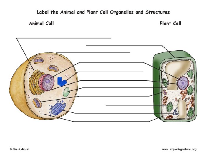
This activity introduces more complex cell structures and incorporates labeling. Provide students with detailed diagrams of both animal and plant cells, including various organelles such as mitochondria, endoplasmic reticulum, Golgi apparatus, vacuoles, and lysosomes. Students will color each organelle using a specific color, and then label each organelle using a key or directly on the diagram. This activity enhances understanding of cell structure and function by requiring students to identify and name different organelles, strengthening their comprehension and recall.
A color-coded key accompanying the diagram simplifies the labeling process and aids understanding.
Creating a Digital Cell Coloring Activity
Digital cell coloring activities can be created using various readily available software. Microsoft PowerPoint or Google Slides allow for the creation of interactive coloring pages where students can digitally “color” the cell structures using the fill tool. More advanced software like Adobe Photoshop or Procreate offers greater control over the design and artistic expression. These programs permit the creation of highly detailed and visually appealing cell diagrams, incorporating layers, textures, and various artistic effects.
The choice of software depends on the desired level of detail and the user’s familiarity with the software. The resulting digital activity can be shared electronically, providing accessibility and flexibility.
Materials Needed for a Hands-on Cell Coloring Activity
A successful hands-on cell coloring activity requires a few key materials. Preparation ensures a smooth and enjoyable learning experience for students.
Understanding the differences between animal and plant cell coloring pdfs can be a fun and educational activity. For instance, the detailed structures within each cell type offer a fascinating contrast. To further explore the animal kingdom, you might also enjoy animal alphabet v coloring pages printable , which provides a different perspective on animal structures. Returning to the cellular level, remember to pay close attention to the key organelles when completing your animal and plant cell coloring pdfs.
- Printed copies of cell diagrams (animal and plant cells)
- Colored pencils, crayons, or markers
- Rulers (optional, for precise labeling)
- Labels or stickers (for the advanced labeling activity)
- Optional: Scissors and glue (for creating a cell model from the coloring page)
Illustrative Examples of Cell Coloring: Animal And Plant Cell Coloring Pdf

Creating visually engaging and informative cell diagrams for coloring requires careful consideration of both accuracy and aesthetic appeal. The following examples illustrate how to effectively depict plant and animal cells, highlighting key organelles and suggesting various coloring styles.
Plant Cell Diagram: Focusing on Chloroplast and Cell Wall
A suitable plant cell diagram for coloring should prominently feature the rigid cell wall, a defining characteristic of plant cells, depicted as a thick, rectangular outer boundary. The cell wall could be colored a light brown or green to represent its composition of cellulose. Within the cell wall, the cell membrane should be clearly visible, a thinner line just inside the cell wall, perhaps in a lighter shade of the cell wall color.
The large central vacuole should be shown occupying a significant portion of the cell’s interior, and could be colored a light purple or blue to represent the watery solution it contains. Crucially, numerous chloroplasts, the sites of photosynthesis, should be depicted as numerous oval or disc-shaped structures scattered throughout the cytoplasm. These chloroplasts should be colored a vibrant green, perhaps with variations in shading to suggest three-dimensionality.
The nucleus, smaller than the vacuole but still prominent, can be shown as a circular structure near the cell membrane, colored a light pink or a pale yellow. Other organelles, like the mitochondria, can be included but less prominently than the chloroplast and cell wall.
Animal Cell Diagram: Highlighting Nucleus and Mitochondria, Animal and plant cell coloring pdf
An effective animal cell diagram for coloring should emphasize the nucleus and mitochondria. The nucleus, the control center of the cell, should be depicted as a large, centrally located, roughly spherical structure. It can be colored a darker pink or a deep purple to contrast with the surrounding cytoplasm. The nuclear membrane, a double membrane surrounding the nucleus, should be a slightly lighter shade of the same color, creating a visual separation.
Within the nucleus, the nucleolus can be indicated as a smaller, darker circle. The mitochondria, the powerhouses of the cell, should be represented as numerous smaller, bean-shaped structures scattered throughout the cytoplasm. These can be colored a deep red or a dark orange to represent their energy-producing role. The cytoplasm itself can be a light beige or pale yellow, allowing the other organelles to stand out clearly.
The cell membrane, the outer boundary of the cell, should be shown as a thin, continuous line, possibly a light blue or grey, to visually differentiate it from the cytoplasm.
Creating Visually Appealing Cell Diagrams
Clarity and accuracy are paramount when creating cell diagrams for coloring. Organelles should be clearly labeled with their names, using a font that is easy to read but not overwhelming. Use consistent shapes and sizes for each organelle to avoid confusion. The diagram should be appropriately scaled, ensuring that the relative sizes of organelles are accurately reflected.
A simple color key can greatly enhance understanding, associating each color with a specific organelle. Consider using a clean, uncluttered background to avoid distracting from the cellular structures. Avoid overlapping organelles excessively; maintaining clear visual separation will improve the educational value.
Examples of Different Coloring Styles
Realistic coloring aims for accurate representation of the organelles’ appearance, using colors and shading that reflect their actual structure and function. A cartoonish style might employ brighter, more saturated colors and simplified shapes, making the diagram more visually engaging for younger audiences. A stylized approach could combine elements of both, using realistic colors but simplified shapes for easier coloring.
For instance, the endoplasmic reticulum could be represented realistically as a network of interconnected tubules in a light blue, while a cartoonish style might simplify it into a series of interconnected loops.
Visual Description of a Cell Diagram Showing the Endoplasmic Reticulum and Golgi Apparatus
Imagine a cell diagram where the endoplasmic reticulum (ER) is depicted as a network of interconnected, flattened sacs and tubules extending throughout the cytoplasm. The rough ER, studded with ribosomes, is represented in a slightly darker shade of blue, while the smooth ER, lacking ribosomes, is shown in a lighter shade of blue. The Golgi apparatus, close to the ER, is depicted as a stack of flattened, membrane-bound sacs, resembling a stack of pancakes.
The Golgi apparatus could be colored a light orange or yellow-brown. The vesicles, small membrane-bound sacs that bud off from the Golgi, are smaller and round, colored a similar shade to the Golgi, but slightly lighter. The spatial relationship between the ER and the Golgi should be clearly shown, with vesicles appearing to transport materials between the two organelles.
Ribosomes, tiny dots on the rough ER, could be colored dark purple or grey.
Detailed FAQs
Where can I find printable versions of the coloring pages?
The PDF will be available for download [link to PDF would go here].
Are there coloring pages suitable for different age groups?
Yes, the guide includes instructions for creating activities suitable for elementary school students and more advanced activities for older learners.
What software can I use to create digital coloring activities?
Many readily available programs, such as Adobe Illustrator or even simple drawing programs, can be used. The guide offers suggestions.
How can I assess student learning from these activities?
The guide includes strategies for assessing understanding, including checking for accurate labeling of organelles and comprehension of biological processes.


