Introduction to Animal Cell Structure: Animal Cell Diagram Coloring Sheet
Animal cell diagram coloring sheet – Embark on a journey into the microcosm, a realm of breathtaking complexity and exquisite design. The animal cell, a fundamental building block of life, reveals a universe of intricate processes within its seemingly simple form. Understanding its structure is akin to understanding the very essence of life itself – a symphony of coordinated activity, a testament to the elegant efficiency of nature’s design.
This exploration will illuminate the key components of this cellular marvel and their profound interconnectedness.The animal cell, unlike its plant counterpart, lacks a rigid cell wall, allowing for greater flexibility and adaptability. This inherent plasticity enables diverse cellular functions and shapes, reflecting the remarkable versatility of life forms. Within this dynamic environment, a multitude of organelles perform their specialized roles, contributing to the overall health and function of the organism.
Each organelle, a miniature powerhouse, is a testament to the intricate design inherent in biological systems.
Understanding animal cell structures can be engaging, especially for younger learners. An animal cell diagram coloring sheet provides a hands-on approach to learning about organelles like the nucleus and mitochondria. To complement this educational activity, consider incorporating fun elements like those found in a animal coloring page printable , which can help maintain interest and reinforce learning.
Returning to the cell diagram, remember to carefully color-code each organelle for better comprehension.
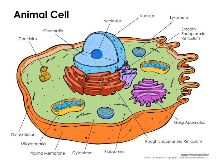
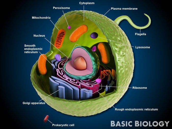
Animal Cell Organelles and Their Functions, Animal cell diagram coloring sheet
The animal cell’s inner workings are a testament to the power of collaboration. Numerous specialized structures, or organelles, work in concert to maintain cellular integrity and carry out essential life processes. These organelles are not isolated entities but rather interconnected components of a finely tuned system. Their coordinated actions are essential for the cell’s survival and function.
- Nucleus: The control center, housing the cell’s genetic material (DNA) and directing cellular activities.
- Ribosomes: Tiny protein factories responsible for synthesizing proteins, the workhorses of the cell.
- Endoplasmic Reticulum (ER): A network of membranes involved in protein and lipid synthesis and transport. The rough ER, studded with ribosomes, is involved in protein synthesis, while the smooth ER plays a role in lipid metabolism and detoxification.
- Golgi Apparatus (Golgi Body): The cell’s packaging and distribution center, modifying, sorting, and transporting proteins and lipids.
- Mitochondria: The powerhouses of the cell, generating energy (ATP) through cellular respiration.
- Lysosomes: The cell’s recycling centers, containing enzymes that break down waste materials and cellular debris.
- Vacuoles: Storage compartments for water, nutrients, and waste products. While smaller and more numerous than in plant cells, they still play a crucial role in maintaining cellular balance.
- Cytoskeleton: A network of protein filaments providing structural support and facilitating intracellular transport.
- Cell Membrane: The outer boundary of the cell, regulating the passage of substances into and out of the cell.
Plant Cell versus Animal Cell: Key Structural Differences
While both plant and animal cells share fundamental similarities, key structural differences highlight their distinct evolutionary paths and functional adaptations. These differences reflect the diverse lifestyles and environmental demands of plants and animals. Understanding these distinctions offers a deeper appreciation for the remarkable diversity of life.The most striking difference lies in the presence of a rigid cell wall in plant cells, absent in animal cells.
This cell wall provides structural support and protection, contributing to the plant’s ability to withstand environmental stresses. Plant cells also possess large, central vacuoles that play a crucial role in maintaining turgor pressure, contributing to the plant’s structural integrity and overall health. Chloroplasts, responsible for photosynthesis, are another defining feature of plant cells, absent in animal cells.
This difference reflects the fundamental distinction between autotrophic (plant) and heterotrophic (animal) modes of nutrition. The presence or absence of these structures directly impacts the cell’s overall structure, function, and interaction with its environment.
Creating a Coloring Sheet Design

Embark on a journey of cellular discovery, where the seemingly simple act of coloring reveals the profound beauty and intricate workings of life itself. This coloring sheet design aims not just to entertain, but to illuminate the inner sanctum of the animal cell, fostering a deeper appreciation for the miraculous machinery within each living being. We will craft a visual representation that is both aesthetically pleasing and scientifically accurate, a harmonious blend of art and science.Consider the animal cell as a vibrant microcosm, a bustling city teeming with specialized structures, each performing its unique function in the grand symphony of life.
Our design will capture this essence, arranging the organelles in a manner that is both visually appealing and intuitively understandable, allowing the colorist to engage with the cell’s architecture in a meaningful way.
Organelle Placement and Size
The nucleus, the cell’s control center, should be positioned centrally and drawn larger than other organelles to reflect its importance. Imagine it as the sun, radiating its influence across the entire cellular landscape. Its size should be approximately one-fifth to one-fourth the diameter of the entire cell. The nucleolus, a smaller, denser region within the nucleus, can be depicted as a smaller circle within the larger nucleus, representing the site of ribosome production.The mitochondria, the cell’s powerhouses, can be depicted as numerous, bean-shaped structures scattered throughout the cytoplasm.
Their size should be relatively small compared to the nucleus, but larger than ribosomes, reflecting their vital role in energy production. Think of them as miniature power plants, fueling the cell’s activities.The endoplasmic reticulum (ER), a network of interconnected membranes, can be represented as a series of interconnected tubes and sacs, extending throughout the cytoplasm. The rough ER, studded with ribosomes, can be distinguished from the smooth ER by depicting small dots (ribosomes) on its surface.
The ER’s size and extent should reflect its role in protein synthesis and lipid metabolism, spanning a significant portion of the cell’s interior.The Golgi apparatus, the cell’s packaging and distribution center, can be illustrated as a stack of flattened sacs, near the nucleus. Its size should be smaller than the nucleus but larger than individual lysosomes. This design mirrors its function in modifying, sorting, and packaging proteins for transport.Lysosomes, the cell’s recycling centers, can be depicted as small, oval-shaped structures scattered throughout the cytoplasm.
Their size should be smaller than the mitochondria, reflecting their role in breaking down waste materials.Finally, the cell membrane, the outer boundary of the cell, should form a complete, smooth Artikel encompassing all the organelles. Its thickness should be relatively thin compared to the internal organelles, highlighting its role as a selective barrier.
Coloring Sheet Instructions and Key

Embark on this artistic journey of cellular exploration, a meditative practice where precision and mindful coloring unveil the intricate beauty of the animal cell. Through careful rendering, we connect with the fundamental building blocks of life, appreciating the divine artistry woven into even the smallest components.This section provides detailed instructions for completing the coloring sheet accurately and a key to match each organelle to its assigned color.
Accurate representation enhances understanding and deepens our appreciation for the cellular world’s harmonious complexity.
Color Suggestions for Organelles
The following list provides a suggested color palette for each organelle, promoting clarity and visual distinction. These choices are merely suggestions; feel free to explore your own creative palette, guided by your inner artistic vision. Remember, the act of coloring itself is a form of meditation, connecting you to the profound beauty of cellular structure.
- Cell Membrane: A soft, translucent blue, representing the boundary that holds the cell’s essence. Imagine the gentle flow of life contained within.
- Cytoplasm: A pale, warm yellow, signifying the life-giving fluid that sustains the cellular processes. Visualize the sun’s nourishing energy within.
- Nucleus: A deep, rich purple, representing the control center, the heart of the cell, where genetic information resides. Imagine the wisdom and potential held within.
- Nucleolus: A vibrant magenta, a smaller, darker shade within the nucleus, highlighting the site of ribosome production. Envision the generative force of life itself.
- Ribosomes: Tiny specks of bright green, scattered throughout the cytoplasm, representing the protein factories. Imagine the industriousness of life’s building blocks.
- Endoplasmic Reticulum (Rough ER): A textured light grey, dotted with green ribosomes, showcasing its role in protein synthesis and transport. Visualize the complex pathways of creation.
- Endoplasmic Reticulum (Smooth ER): A smoother, pale grey, distinct from the rough ER, reflecting its functions in lipid synthesis and detoxification. Imagine the cell’s capacity for transformation and purification.
- Golgi Apparatus: A layered, warm orange, representing the processing and packaging center. Imagine the organization and refinement of cellular products.
- Mitochondria: Lively crimson, signifying the powerhouses of the cell, generating energy. Visualize the dynamic energy that sustains life.
- Lysosomes: A deep, cool blue, representing the waste disposal and recycling centers. Imagine the cell’s capacity for self-renewal and purification.
- Centrioles: Small, dark brown circles near the nucleus, illustrating their role in cell division. Imagine the cell’s potential for growth and renewal.
Color Key
This key provides a clear correspondence between each organelle and its suggested color. Use this as a guide, but remember that the true beauty lies in your personal interpretation and creative expression. Let your intuition guide your color choices, mirroring the vibrant energy of life within.
| Organelle | Suggested Color |
|---|---|
| Cell Membrane | Soft, translucent blue |
| Cytoplasm | Pale, warm yellow |
| Nucleus | Deep, rich purple |
| Nucleolus | Vibrant magenta |
| Ribosomes | Bright green |
| Rough ER | Textured light grey (dotted with green) |
| Smooth ER | Smooth pale grey |
| Golgi Apparatus | Layered warm orange |
| Mitochondria | Lively crimson |
| Lysosomes | Deep, cool blue |
| Centrioles | Dark brown |


